How an FTIR Spectrometer Operates

Applications:-
- organic synthesis,
- polymer science,
- petrochemical engineering,
- pharmaceutical industry and food analysis
Introduction
The range of Infrared region is 12800 ~ 10 cm-1and can be divided into near-infrared region (12800 ~ 4000 cm-1), mid-infrared region (4000 ~ 200 cm-1) and far-infrared region (50 ~ 1000 cm-1).
Infrared absorption spectroscopy is the method which scientists use to determine the structures of molecules with the molecules’ characteristic absorption of infrared radiation. Infrared spectrum is molecular vibrational spectrum. When exposed to infrared radiation, sample molecules selectively absorb radiation of specific wavelengths which causes the change of dipole moment of sample molecules. Consequently, the vibrational energy levels of sample molecules transfer from ground state to excited state. The frequency of the absorption peak is determined by the vibrational energy gap. The number of absorption peaks is related to the number of vibrational freedom of the molecule. The intensity of absorption peaks is related to the change of dipole moment and the possibility of the transition of energy levels. Therefore, by analyzing the infrared spectrum, one can readily obtain abundant structure information of a molecule. Most molecules are infrared active except for several homonuclear diatomic molecules such as O2, N2 and Cl2due to the zero dipole change in the vibration and rotation of these molecules. What makes infrared absorption spectroscopy even more useful is the fact that it is capable to analyze all gas, liquid and solid samples. The common used region for infrared absorption spectroscopy is 4000 ~ 400 cm-1 because the absorption radiation of most organic compounds and inorganic ions is within this region.
FTIR spectrometers are the third generation infrared spectrometer. FTIR spectrometers have several prominent advantages:
FTIR spectrometers are the third generation infrared spectrometer. FTIR spectrometers have several prominent advantages:
- The signal-to-noise ratio of spectrum is significantly higher than the previous generation infrared spectrometers.
- The accuracy of wavenumber is high. The error is within the range of ± 0.01 cm-1.
- The scan time of all frequencies is short (approximately 1 s).
- The resolution is extremely high (0.1 ~ 0.005 cm-1).
- The scan range is wide (1000 ~ 10 cm-1).
- The interference from stray light is reduced. Due to these advantages, FTIR Spectrometers have replaced dispersive IR spectrometers.
The Components of FTIR Spectrometers
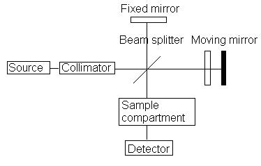
A common FTIR spectrometer consists of a source, interferometer, sample compartment, detector, amplifier, A/D convertor, and a computer. The source generates radiation which passes the sample through the interferometer and reaches the detector. Then the signal is amplified and converted to digital signal by the amplifier and analog-to-digital converter, respectively. Eventually, the signal is transferred to a computer in which Fourier transform is carried out. Figure 2 is a block diagram of an FTIR spectrometer.
 |
| Block diagram of an FTIR spectrometer |
 |
| IR spectrum of a sample |
Operation for FTIR Spectrometer
Step 1: The first step is sample preparation. The standard method to prepare solid sample for FTIR spectrometer is to use KBr. About 2 mg of sample and 200 mg KBr are dried and ground. The particle size should be unified and less than two micrometers. Then, the mixture is squeezed to form transparent pellets which can be measured directly. For liquids with high boiling point or viscous solution, it can be added in between two NaCl pellets. Then the sample is fixed in the cell by skews and measured. For volatile liquid sample, it is dissolved in CS2 or CCl4 to form 10% solution. Then the solution is injected into a liquid cell for measurement. Gas sample needs to be measured in a gas cell with two KBr windows on each side. The gas cell should first be vacuumed. Then the sample can be introduced to the gas cell for measurement.
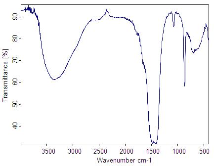
Step 2: The second step is getting a background spectrum by collecting an interferogram and its subsequent conversion to frequency data by inverse Fourier transform. We obtain the background spectrum because the solvent in which we place our sample will have traces of dissolved gases as well as solvent molecules that contribute information that are not our sample. The background spectrum will contain information about the species of gases and solvent molecules, which may then be subtracted away from our sample spectrum in order to gain information about just the sample. Figure 6 shows an example of an FTIR background spectrum.
The background spectrum also takes into account several other factors related to the instrument performance, which includes information about the source, interferometer, detector, and the contribution of ambient water (note the two irregular groups of lines at about 3600 cm–1 and about 1600 cm–1 in Figure 6) and carbon dioxide (note the doublet at 2360 cm–1 and sharp spike at 667 cm–1 inFigure 6) present in the optical bench.
Step 3: Next, we collect a single-beam spectrum of the sample, which will contain absorption bands from the sample as well as the background (gaseous or solvent).
Step 4: The ratio between the single-beam sample spectrum and the single beam background spectrum gives the spectrum of the sample (Figure 7).
|
| Sample IR spectrum |
Outside Links
- http://chemwiki.ucdavis.edu/Physical_Chemistry/Spectroscopy/Vibrational_Spectroscopy/Infrared_Spectroscopy/
- http://infrared.als.lbl.gov/content/web-links
- http://www.fc.up.pt/pessoas/peter.eaton/tutorial/webCT/
- http://www.oceanoptics.com/lol/
- http://www.youtube.com/watch?v=6isp-G4XBB8&feature=related
- http://mmrc.caltech.edu/FTIR/FTIRintro.pdf
- http://www.youtube.com/watch?v=ObklYbQaX24&NR=1&feature=fvwp
- http://mmrc.caltech.edu/FTIR/FTIRintro.pdf
No comments:
Post a Comment